Diphyllobothrium spp.
Contributor: Manon Simard
Biology: Diphyllobothrium spp. are flat, segmented, ribbon like, worms that can reach 10 m long, that can be seen in intestines in which they feed. The head called the scolex is spoon-shaped, measure 1 mm x 2.5 mm and has 2 grooves along its length. The segments are short measuring 2-4mm long and a width of 10-12mm. Eggs are pale brown with an operculum and round extremities. They are 70μm (55-76μm) long and have a width of 50μm (40-60μm). There are at least 15 species in the world but only 6 of them are infectious to humans.
Life cycle: The eggs of the parasite located in a fish are released with the faeces in the water. Eggs will hatch in the water and release larva which are eaten by copepods. The second stage larvae also named plerocercoid, develop into the copepod within 2 to 3 weeks. A third stage larva is formed once a fish eats infected copepods. The parasite will stay in a state of dormancy and will only become an adult until a mammal predator eats infected fish.
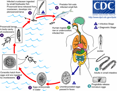
Figure 1. Life cycle of Diphyllobothrium spp (Source: CDC).
Transmission: Transmission occurs during consumption of raw or undercooked, lightly marinated, salted and smoked infected fish. Sushi and sashimi that contains raw fish is also a source of infection. There is more chance to be infected with Diphyllobothrium spp. that are living in the flesh (D. latum) than in the body cavity (D. dendriticum).
Prevalence: There are at least 20 millions of people infected worldwide, and has been reported in all provinces and territories of Canada except the Maritimes.
Susceptible populations: People who eat raw or undercooked fish, poorly smoked and marinated and lightly salted infected fish. Most records of Diphyllobothriasis are in North America (especially Canada), northern Europe, and Far East countries. A few cases are reported in South America, and Uganda. Diphyllobothrium latum has been reported in temperate and sub-arctic regions and D. dendriticum are reported in Arctic regions. However, newly developed identification techniques are used to confirm these reports and update the distribution of each species.
Symptoms: Most humans are asymptomatic however, symptoms may include diarrhea, vomiting, weight loss. Complications may lead to intestinal obstruction and anemia, development of a gall bladder disease caused by proglottid migration.
Treatment: Diagnosis is made by the identification of the eggs or segments of the parasites in stools. Medication such as praziquantel and niclosamides are most often used.
Control Measures: Proper cooking and freezing will control its transmission to humans. Frying or boiling fish at 550°C for 5 minutes or 500°C for 10 minutes will kill the plerocercoids whereas freezing at -100°C for 8 to 72 hours (depending on depthness of flesh) or -200°C for 24 hours, salting at more than 12% NaCl (salt), and dried salting are also efficient. Smoking does not kill the fish. Larval forms can be seen in the flesh with the eye and can be removed. Removing fish organs as soon as possible reduces the risk of infection since the plerocercoids migrate from organs to flesh when the animal is dead.